
Atrial, ventricular, and arterial blood pressure waveforms each provide a unique view into the cardiovascular system’s functioning.
The atrial blood pressure waveform reflects the changes in pressure within the atria of the heart. It consists of three key components: the A-wave, which represents atrial contraction; the C-wave, which occurs when the ventricle contracts and pushes against the atria; and the V-wave, which represents the period when the atria are filling with blood while the valves are closed. These waveforms help assess atrial function and are commonly monitored through central venous or jugular venous pressure readings.
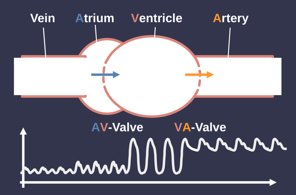
The ventricular blood pressure waveform captures the pressure changes within the heart’s ventricles. It shows a sharp increase during the systolic phase, when the ventricles contract to pump blood into the arteries, and a decrease during the diastolic phase, when the ventricles relax and fill with blood. This waveform is important for understanding how well the ventricles are functioning and can reveal issues like heart failure or valve disorders.
Finally, the arterial blood pressure waveform illustrates the pressure in the arteries as blood is ejected from the heart. The waveform rises sharply during systole when blood is pumped from the left ventricle, and it features a small dip known as the dicrotic notch, which corresponds to the closure of the aortic valve. The waveform then gradually declines during diastole as blood continues to flow through the arteries while the heart is at rest. Arterial waveforms are crucial for monitoring blood pressure and overall cardiovascular health, particularly in critical care settings.
Each waveform provides essential insights into different aspects of heart and circulatory function, aiding in the diagnosis and management of various cardiovascular conditions.