
Echocardiography is a vital diagnostic tool in cardiology, employing high-frequency sound waves to create detailed images of the heart’s structure and function. This non-invasive procedure allows for real-time assessment of the heart without the need for radiation, making it safe for a wide range of patients.
The process begins with the application of a gel on the chest to facilitate sound wave transmission. A transducer, which emits and receives sound waves, is then placed on the skin. As the sound waves travel through the body, they encounter different tissues and structures, producing echoes. These echoes are captured by the transducer and converted into images displayed on a monitor.
There are several types of echocardiography, including transthoracic echocardiography (TTE) and transesophageal echocardiography (TEE). TTE is the most common, where the transducer is positioned on the chest wall. TEE, on the other hand, involves placing the transducer in the esophagus for a closer view of the heart, providing more detailed images, especially in cases where TTE is insufficient.
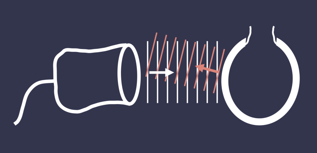
Echocardiography can assess various aspects of heart health, such as chamber sizes, wall motion, valve function, and blood flow patterns. Doppler echocardiography, a specialized technique, measures the speed and direction of blood flow, aiding in the evaluation of conditions like valve stenosis or regurgitation.
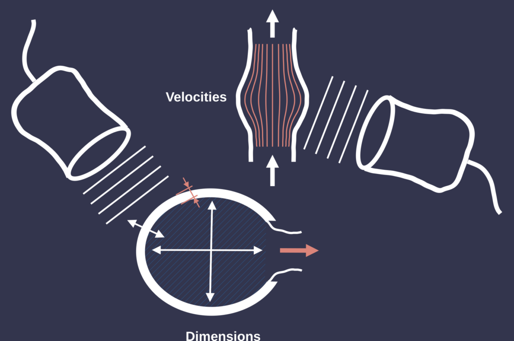
Echocardiography is capable of measuring several key variables essential for assessing heart function and structure. One of the primary metrics is chamber size, where the dimensions of the heart’s atria and ventricles are evaluated to identify any enlargement or abnormal sizing. Additionally, wall motion is analyzed to observe the movement of the heart walls during contraction, helping to detect any regional wall motion defects that may indicate ischemia.
Another critical variable is the ejection fraction, which represents the percentage of blood the left ventricle pumps out with each contraction. This measurement is vital for assessing overall heart function. Echocardiography also evaluates valve function by examining the structure and movement of the heart valves, allowing clinicians to assess conditions such as stenosis or regurgitation.
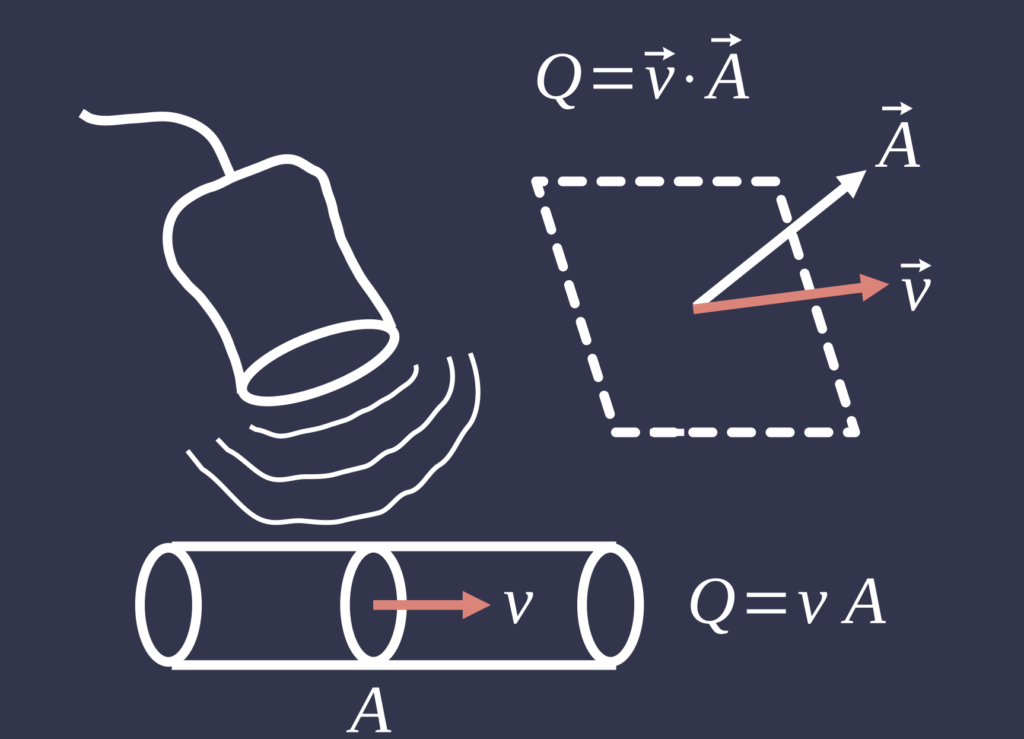
Doppler imaging is employed to measure blood flow, capturing the speed and direction of blood as it moves through the heart and vessels. This helps identify abnormalities in blood flow patterns. Additionally, echocardiography can detect congenital defects, such as atrial or ventricular septal defects, by visualizing the flow of blood between heart chambers.
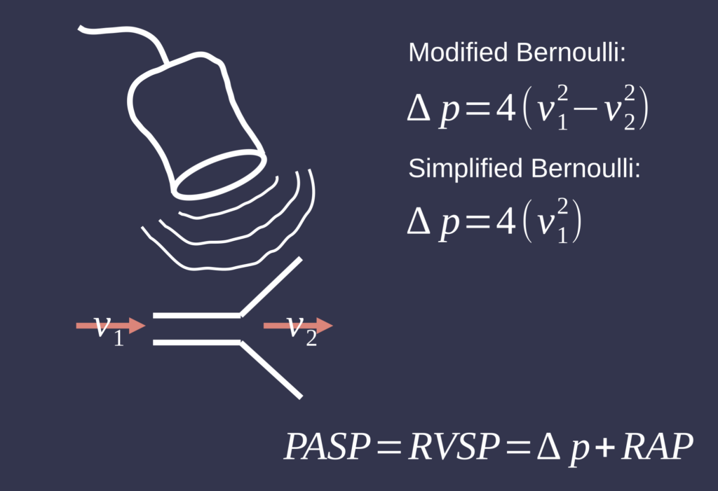
By analyzing these blood flow velocities, they can estimate pressure gradients across the heart’s chambers and valves using the Bernoulli principle. The resulting images and flow data are displayed on a monitor, allowing for a comprehensive assessment of cardiac function and potential valve abnormalities.
Finally, the presence of pericardial effusion—fluid accumulation around the heart—can be identified, which may indicate various underlying conditions. Together, these measurements provide comprehensive insights into cardiac health, playing a crucial role in diagnosis and treatment planning.
While echocardiography is a valuable tool, it does have some limitations and potential inaccuracies. Factors such as patient body habitus, poor acoustic windows due to lung disease or obesity, and operator skill can affect image quality and measurement accuracy. Additionally, subtle abnormalities, particularly in complex cases, might be missed or misinterpreted. Doppler measurements can also be influenced by angle dependence, which may lead to errors in assessing blood flow velocities. Despite these challenges, echocardiography remains a cornerstone in cardiac evaluation, often supplemented by other diagnostic methods for more comprehensive assessments.